Accepted at: Aug, 22, 2024
4:13 a.m.
Author:
Solar04
Related Deck:
1680573171560
Accepted
Rationale for new note
Updating notes on topic. As previous notes were outdated.
Text
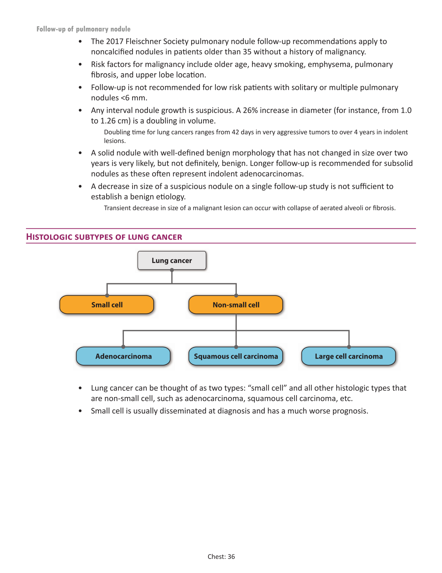
Thoracic Imaging - Lung cancer
The Fleischner Society pulmonary nodule follow-up recommendations:
SUBSOLID NODULES
The Fleischner Society pulmonary nodule follow-up recommendations:
SUBSOLID NODULES
Multiple subsolid nodules <6 mm (<100 mm3)
- {{c1::CT at 3-6 months, then if stable consider CT at 2 and 4 years in high-risk patients::f/u?}}
- {{c2::CT at 3-6 months, then subsequent management based on the most suspicious nodule(s)::f/u?}}
Text
<i>Thoracic Imaging - Lung cancer<br></i><br>The <strong>Fleischner Society pulmonary nodule follow-up recommendations:<br></strong><br>SUBSOLID NODULES<br><br><div>Multiple subsolid nodules <6 mm <strong>(</strong><100 mm<sup>3</sup>)<ul><li><div>{{c1::CT at 3-6 months, then if stable consider CT at 2 and 4 years in high-risk patients::f/u?}}</div></li></ul>Multiple subsolid nodules ≥6 mm (>100 mm<sup>3</sup>)<ul><li><div>{{c2::CT at 3-6 months, then subsequent management based on the most suspicious nodule(s)::f/u?}}</div></li></ul></div>
Extra
Solid nodules
Source: Radiopedia Fleischner Society pulmonary nodule recommendations | Radiology Reference Article | Radiopaedia.org
The Fleischner Society pulmonary nodule recommendations pertain to the follow-up and management of indeterminate pulmonary nodules detected incidentally on CT and are published by the Fleischner Society. The guideline does not apply to lung cancer screening, patients younger than 35 years, or patients with a history of primary cancer or immunosuppression.
NB: This article reflects the 2017 revision 4, which supersedes prior versions published in 2005 1 and 2013 2.
Solid nodules
Single
Single solid nodule <6 mm (<100 mm3)
- low-risk patients: no routine follow-up required
- high-risk patients: optional CT at 12 months (particularly with suspicious nodule morphology and/or upper lobe location; see "risk assessment" below)
- low-risk patients: CT at 6-12 months, then consider CT at 18-24 months
- high-risk patients: CT at 6-12 months, then CT at 18-24 months
- low-risk and high-risk patients: consider CT at 3 months, PET-CT, or tissue sampling
Multiple solid nodules <6 mm (<100 mm3)
- low-risk patients: no routine follow-up required
- high-risk patients: optional CT at 12 months
- low-risk patients: CT at 3-6 months, then consider CT at 18-24 months
- high-risk patients: CT at 3-6 months, then CT at 18-24 months
When multiple nodules are present, the most suspicious nodule should guide further individualized management.
Subsolid nodulesSingle
Single ground glass nodule <6 mm (<100 mm3)
- no routine follow-up required
- CT at 6-12 months, then if persistent, CT every 2 years until 5 years
- CT at 3-6 months, then if persistent and solid component remains <6 mm, annual CT until 5 years
Multiple subsolid nodules <6 mm (<100 mm3)
- CT at 3-6 months, then if stable consider CT at 2 and 4 years in high-risk patients
- CT at 3-6 months, then subsequent management based on the most suspicious nodule(s)
Extra
<div> <div> <div><div><i>Source: Radiopedia <a href="https://radiopaedia.org/articles/fleischner-society-pulmonary-nodule-recommendations-1?lang=us">Fleischner Society pulmonary nodule recommendations | Radiology Reference Article | Radiopaedia.org</a></i></div><br><div>The <strong>Fleischner Society pulmonary nodule recommendations</strong> pertain to the follow-up and management of indeterminate <a href="https://radiopaedia.org/articles/pulmonary-nodule-1?lang=us">pulmonary nodules</a> detected incidentally on CT and are published by the <a href="https://radiopaedia.org/articles/fleischner-society?lang=us">Fleischner Society</a>. The guideline does not apply to lung cancer screening, patients younger than 35 years, or patients with a history of primary cancer or immunosuppression.</div><div>NB: This article reflects the 2017 revision <sup>4</sup>, which supersedes prior versions published in 2005 <sup>1 </sup>and 2013 <sup>2</sup>.</div><br>Solid nodules</div><div>Single</div><div>Single solid nodule <6 mm (<100 mm<sup>3</sup>)<ul><li><div>low-risk patients: no routine follow-up required</div></li><li><div>high-risk patients: optional CT at 12 months (particularly with suspicious nodule morphology and/or upper lobe location; see "risk assessment" below)</div></li></ul>Solitary solid nodule 6-8 mm (100-250 mm<sup>3</sup>)<ul><li><div>low-risk patients: CT at 6-12 months, then consider CT at 18-24 months</div></li><li><div>high-risk patients: CT at 6-12 months, then CT at 18-24 months</div></li></ul>Solitary solid nodule >8 mm (>250 mm<sup>3</sup>)<ul><li><div>low-risk and high-risk patients: consider CT at 3 months, PET-CT, or tissue sampling</div></li></ul>Multiple</div><br><div>Multiple solid nodules <6 mm (<100 mm<sup>3</sup>)<ul><li><div>low-risk patients: no routine follow-up required</div></li><li><div>high-risk patients: optional CT at 12 months</div></li></ul>Multiple solid nodules >6 mm (>100 mm<sup>3</sup>)<ul><li><div>low-risk patients: CT at 3-6 months, then consider CT at 18-24 months</div></li><li><div>high-risk patients: CT at 3-6 months, then CT at 18-24 months</div></li></ul><div>When multiple nodules are present, the most suspicious nodule should guide further individualized management.</div>Subsolid nodules</div><div>Single</div><div>Single ground glass nodule <6 mm (<100 mm<sup>3</sup>)<ul><li><div>no routine follow-up required</div></li></ul>Single ground glass nodule ≥6 mm (>100 mm<sup>3</sup>)<ul><li><div>CT at 6-12 months, then if persistent, CT every 2 years until 5 years</div></li></ul>Single part-solid nodule ≥6 mm (>100 mm<sup>3</sup>)<ul><li><div>CT at 3-6 months, then if persistent and solid component remains <6 mm, annual CT until 5 years</div></li></ul>Multiple</div><div>Multiple subsolid nodules <6 mm <strong>(</strong><100 mm<sup>3</sup>)<ul><li><div>CT at 3-6 months, then if stable consider CT at 2 and 4 years in high-risk patients</div></li></ul>Multiple subsolid nodules ≥6 mm (>100 mm<sup>3</sup>)<ul><li><div>CT at 3-6 months, then subsequent management based on the most suspicious nodule(s)</div></li></ul></div> </div> </div>
Personal Notes
Empty field
Personal Notes
Missed Questions
Empty field
Missed Questions
Core Radiology
Core Radiology
<img src="0036_2E.png">
Fundamentals of Diagnostic Radiology
Empty field
Fundamentals of Diagnostic Radiology
Crack the Core
Empty field
Crack the Core
War Machine
Empty field
War Machine
Sectional Anatomy
Empty field
Sectional Anatomy
Additional Resources
Empty field
Additional Resources
One by one
Empty field
One by one
Section Title
Empty field
Section Title
Tags
#Ankore::Textbooks::Core_Radiology::0036
AnkiHub_Subdeck::AnKore_for_ABR_Diagnostic_Radiology_Core_Exam::Core_Radiology::01_Thoracic_Imaging::05_Lung_cancer
Core_Radiology_2E